
There are essentials tasks that every winemaker should be able to do with a microscope. First you should be able to distinguish between bacteria, yeast, and fungi under the microscope. Second you should be able to tell living organisms from debris (plant cells, crystals, filter and fining agents, etc.). Third you should be able to identify the most common and easily identifiable organisms by sight so that you can tell when you are heading for a problem. With some experience you will know enough to tell that you don’t know what is growing in the wine and take action or get help quickly. There are images of the most common of the wine yeast, bacteria, and mold with their descriptions on this web site. Lastly you should be able to count yeast cells and distinguish between live and dead cells. These are some of the goals we strive for in the Wine Microbiology lab class taught at UC Davis.
Types of microscopy: There are different types of microscopes available ranging in price from a few hundred dollars to many thousands of dollars. Typically for a winery a good quality optical scope is desirable, one with phase contrast is helpful when looking at bacteria. Most microscopes have 10x oculars and objectives from 10x to 100x. This will give you a range of magnification from 100x to 1000x. Sometimes you can get oculars that are 15x or 20x that can increase your magnification without having to use a 100x objective. Any 100x objective requires oil (oil immersion) to decrease the refraction from the light traveling through air between the sample and the objective. This effectively increases the resolution of objects under the microscope. A 20x ocular with a 40x objective will give you a total magnification of 800x without the use of oil immersion. However the resolution is not as good. Besides objectives at 10x, 40x, and 100x some scopes may have 4x, 16x, or 20x objectives as well. The figure below shows Saccharomyces cerevisiae visualized at different magnifications (100x, 400x, 1000x).
Saccharomyces cerevisiae

|
|
|
There are now very inexpensive digital microscopes on the market that replace the ocular with a digital screen similar to what you find in digital cameras. The objectives are the same as a standard microscope but some people find it easier to view the screen than look through the oculars. The digital scope allows for a digital zoom of up to 4x but no optical zoom. The digital zoom can result in pixilation of the image. As with any microscope the quality of the optics determines the quality of the image and most digital microscopes do not have the best quality optics. The best optical microscopes now have a third optical port for the insertion of a digital camera lens. This allows images to be viewed on a computer screen. Use of a digital camera makes it easy to share images with colleagues and some users find the screen easier to use than the traditional ocular.
There are also different types of microscopy for specific purposes. The one you are most familiar with is probably bright field microscopy, which uses light to illuminate an object that absorbs light in denser areas to give contrast for visualization. Dark field microscopy excludes the non-scattered light from the field of view leaving the background dark and the object in the field of view light. Fluorescent microscopy uses a mercury lamp and a filter to illuminate the viewing field with light of a specific wavelength. A second filter filters out that wavelength but allows the light emitted from an excited molecule to pass through the ocular and be visualized. Many biological specimens, especially photosynthetic ones, have a great deal of natural fluorescence, but more typically samples are stained or genetically tagged with fluorescent dyes or labels to allow visualization of structures or specific compounds in cells. Phase contrast and differential interference contrast (DIC) microscopy are techniques that are used to enhance the contrast of low contrast objects especially in unfixed specimens. Use of a phase contrast or DIC microscope for viewing bacteria makes it far easier for an inexperienced user to visualize small, low contrast bacteria. In phase contrast microscopy phase shifts in light are converted to brightness in the field of view allowing normally transparent structures to be seen. DIC works in a similar fashion but uses interferometry rather than phase shifts to allow visualization of invisible structures in cells. Phase contrast is more common and less expensive than DIC but DIC visualized samples have less light halos than phase contrast samples. Below are photographs of Oenococcus oeniusing bright field at 400x, phase contrast, and DIC at 1000x.
Oenococcus oeni
|
|
|
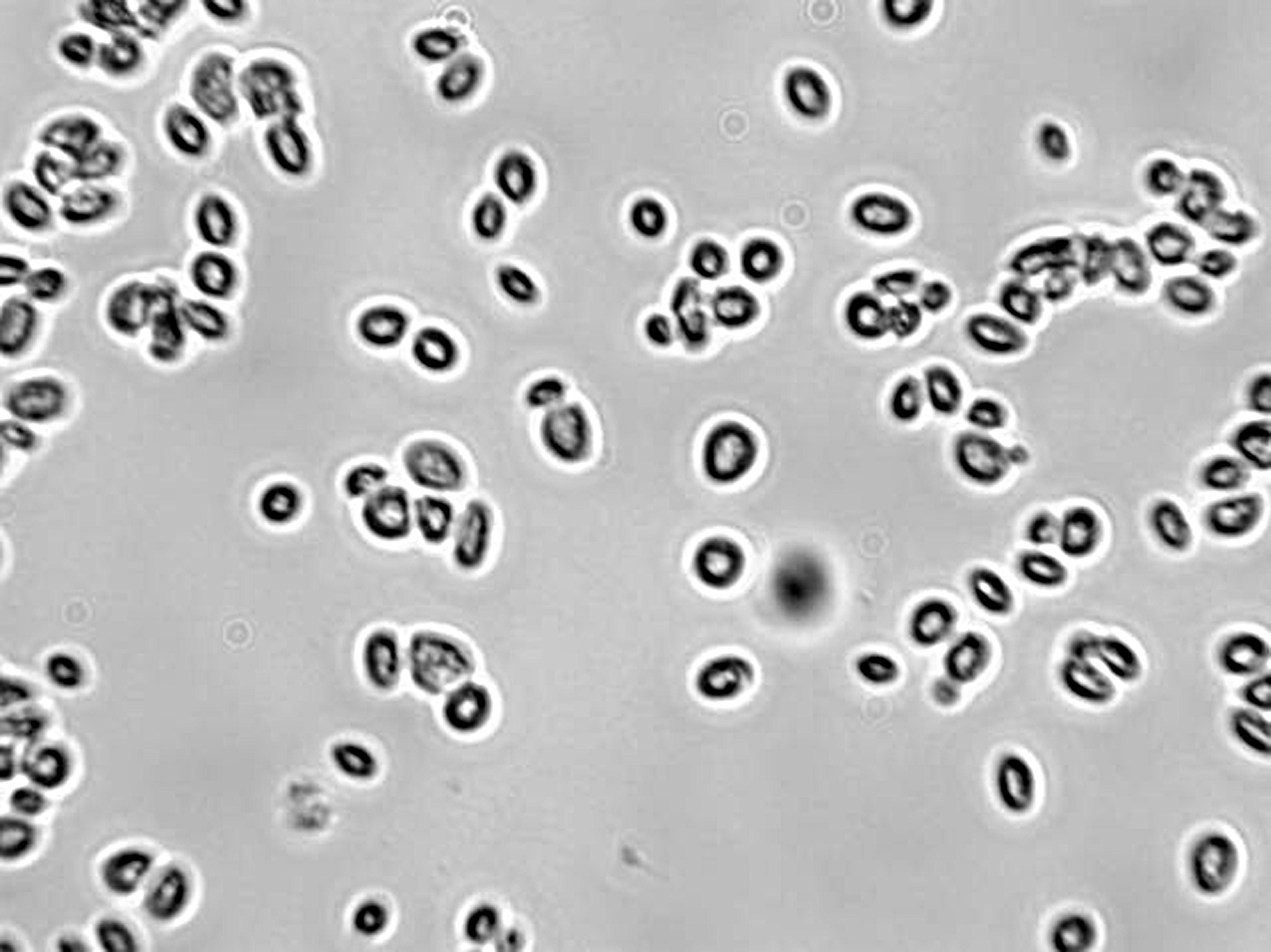
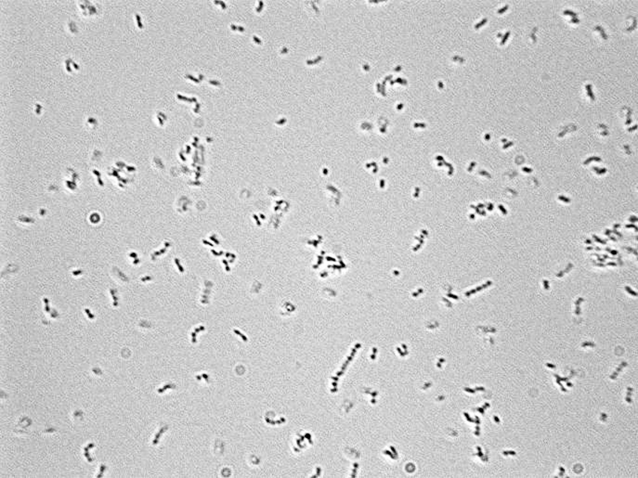
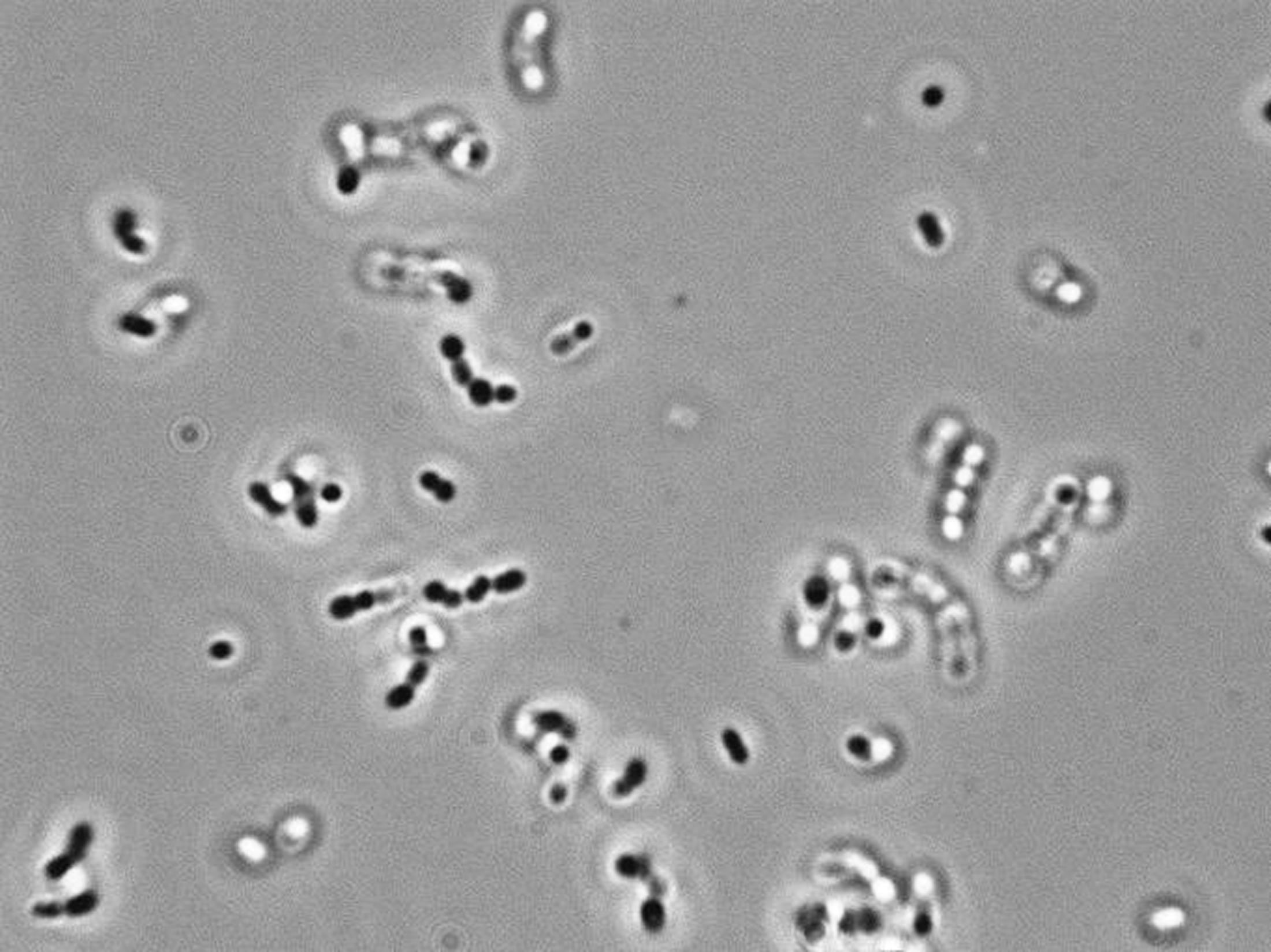
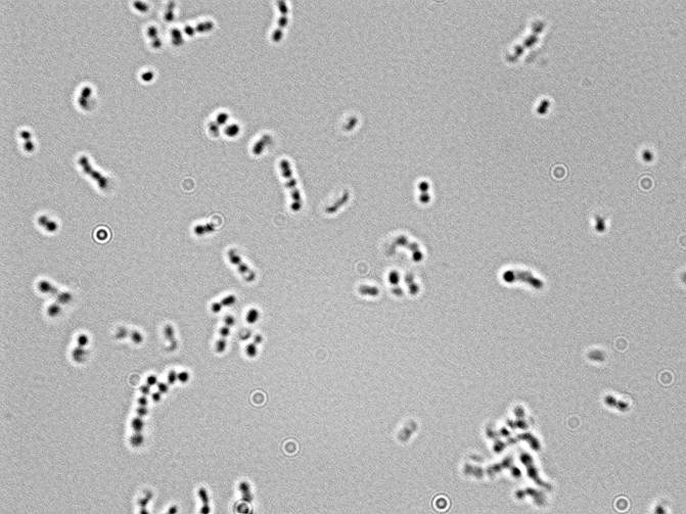
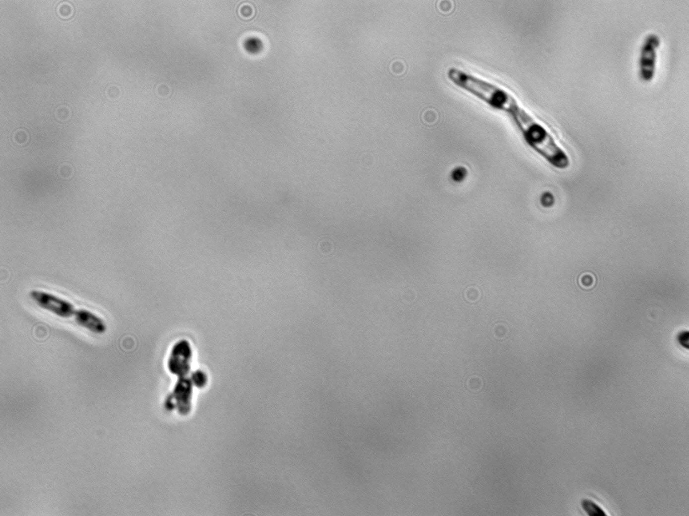
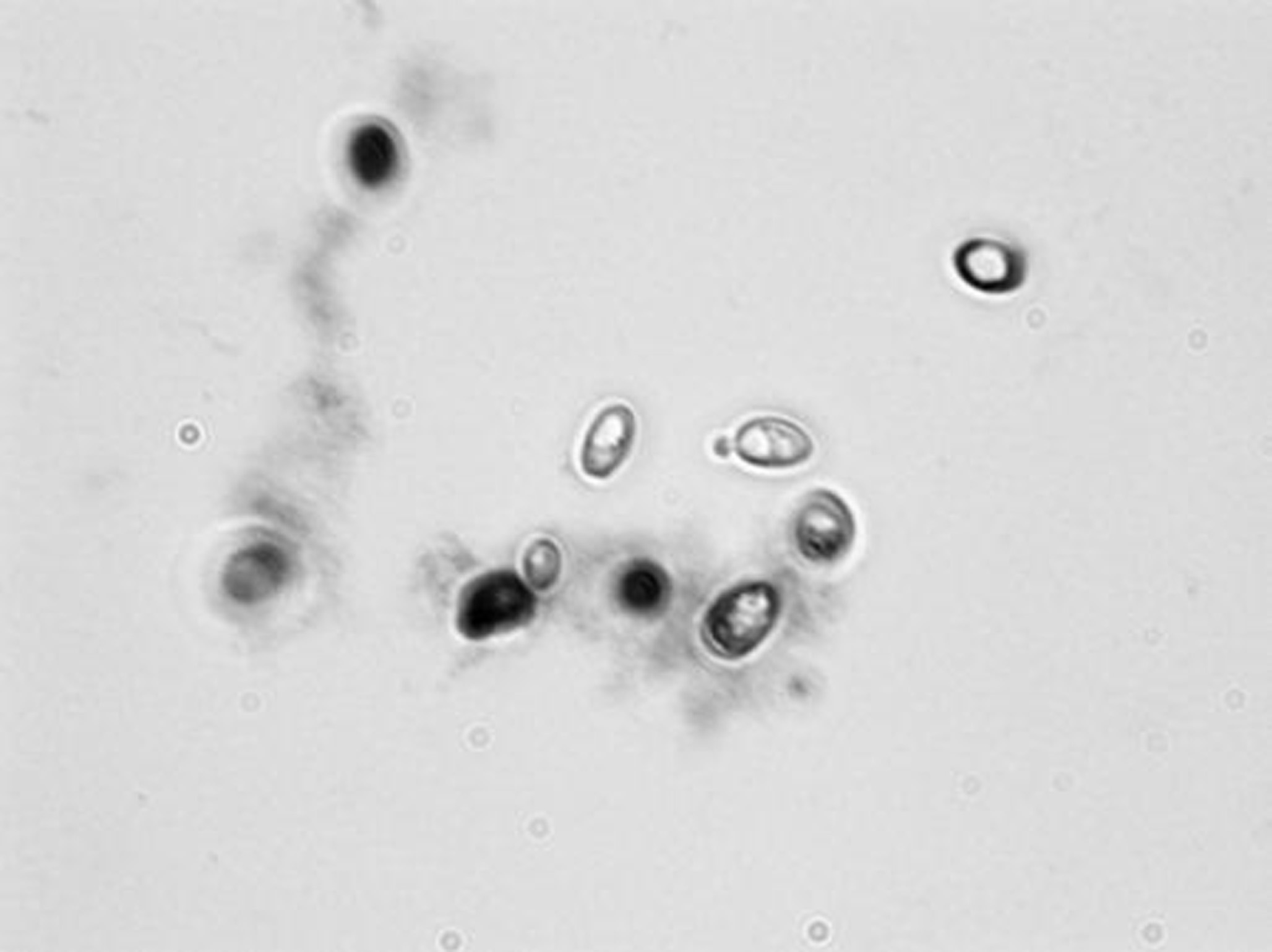
Differentiating yeast, bacteria, and mold: The easiest way to differentiate bacteria, yeast (single celled fungi), and mold (filamentous fungi) is generally by size. Molds are easy to see at 100x magnification, yeast at 400x magnification, and bacteria are usually hard to see unless you go to 1000x magnification. However comparing the size of these organisms can be difficult without a reference. It is often easier if you mix cultures on a single microscope slide or if they are already mixed in a fermentation sample. But there are some commonly seen organisms in the winery that are not as easy to distinguish. Below is a mixture of the common soil bacterium Bacillus megaterium and a Lactobacillus from a wine sample. Bacillus megateriumis the most common non-wine Bacillus that we find in California wines and as its name implies it is a very large bacterium.
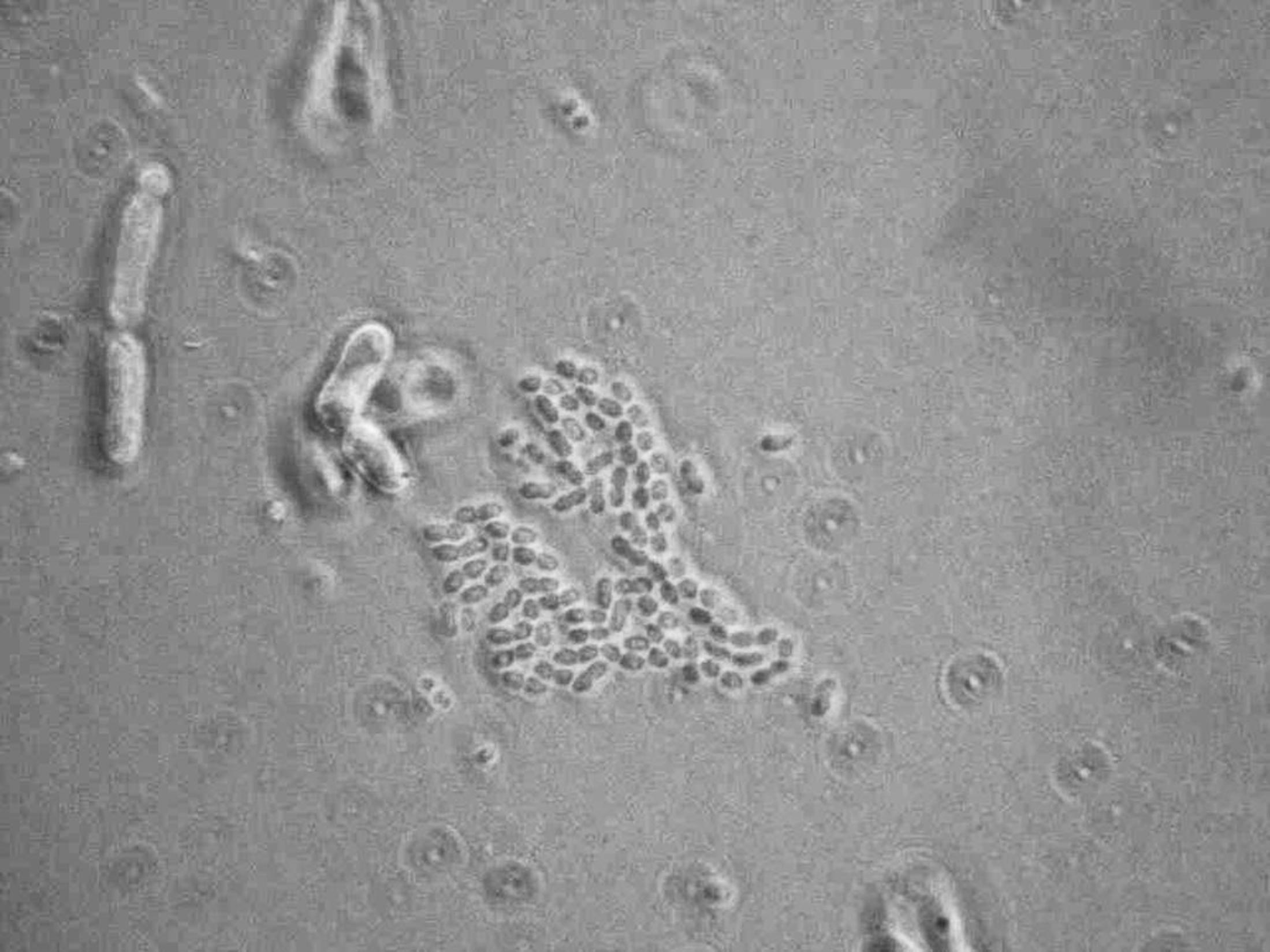
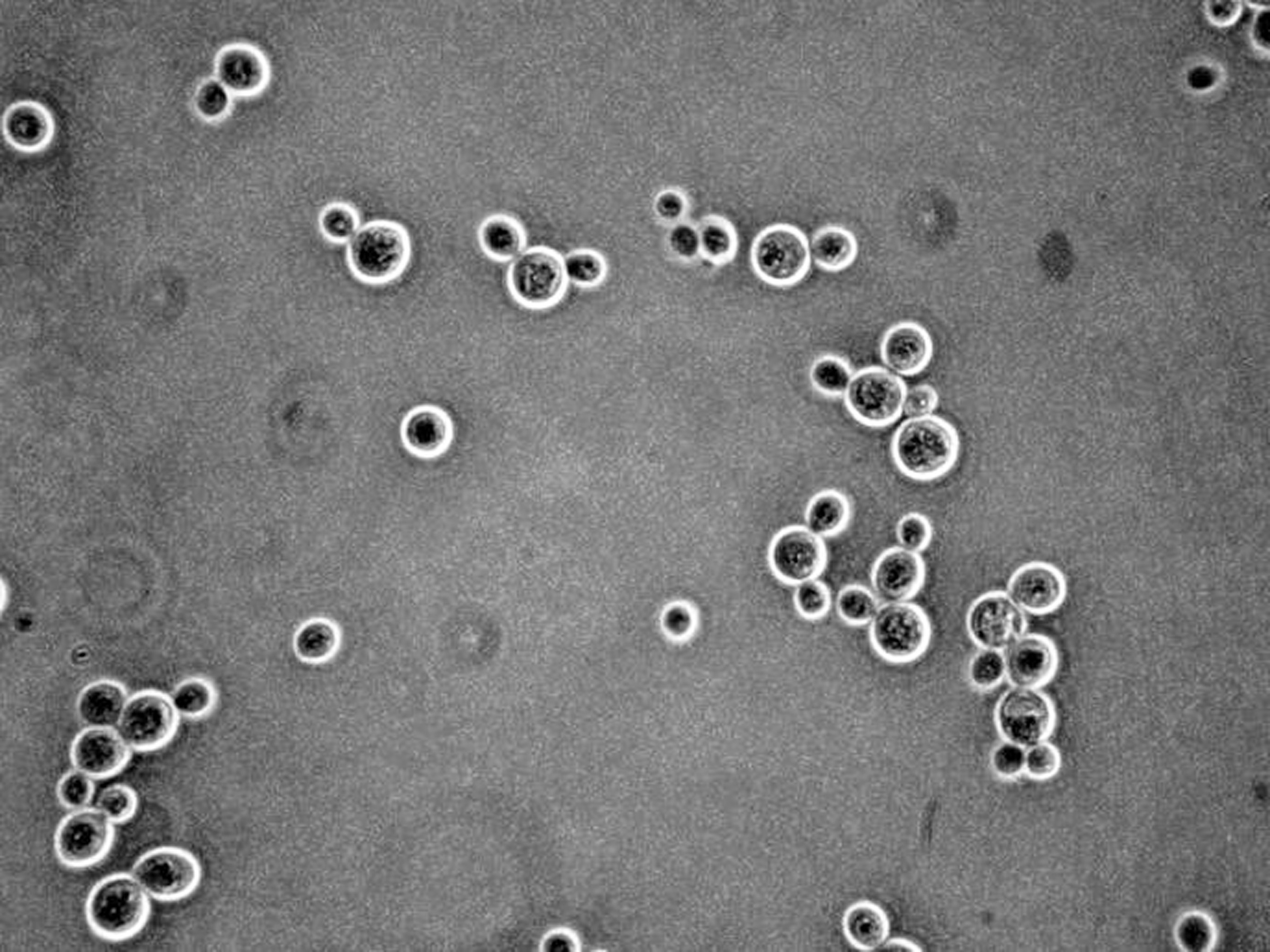
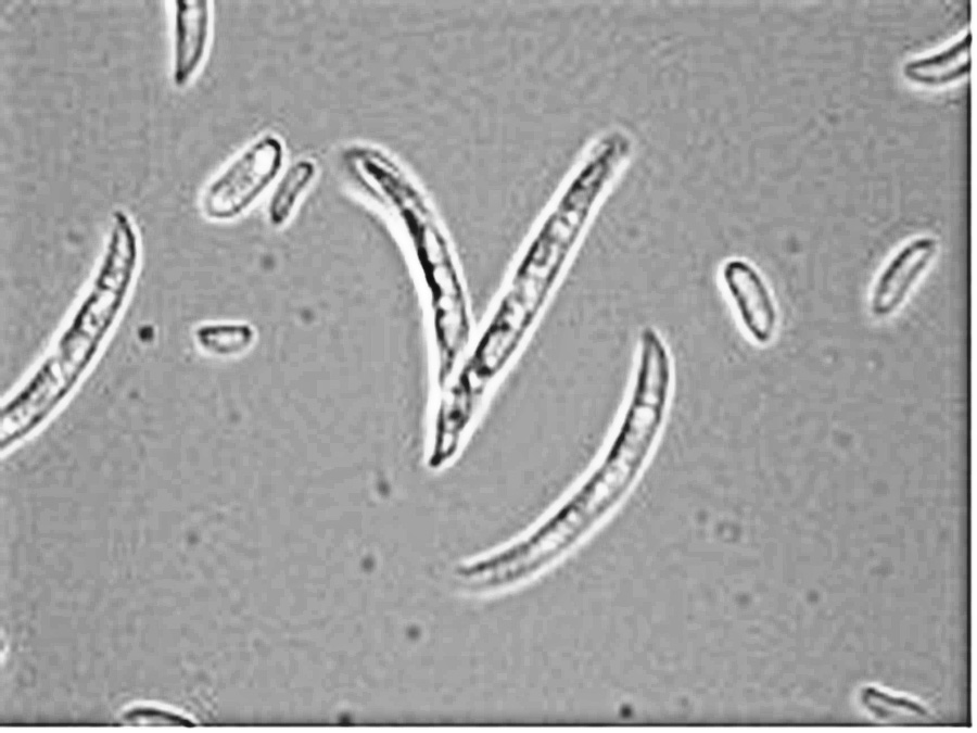
While bacteria are usually much smaller than yeast, Bacillus megaterium is much larger than most bacteria. In the image below the B. megaterium is seen in comparison to Saccharomyces cerevisiae. Even though the sizes are similar, the bacteria are less refractile, more transparent and difficult to see. This is also typical of bacteria versus yeast. The second image is of a Schizosaccharomyces yeast which divides by fission rather then budding. Because bacteria also divide by fission, a large bacterium, like B. megaterium, could be mistaken for a fission yeast. In the case below both images are at the same magnification and you can see that the B. megaterium is smaller than the yeasts.
|
|
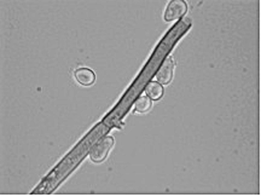
Yeasts are also smaller then molds, which are filamentous, whereas yeasts are single celled. Usually this makes them easy to tell apart but there are exceptions. Most importantly in a wine environment are yeasts, such as Brettanomyces bruxellensis, that form pseudohyphae. Below is a direct comparison of pseudohyphae from Brettanomyces and hyphae fromBotrytis cinerea. The first panel is Brettanomyces at 1000x magnification and the second is Botrytis at 100x magnification. TheBotrytis is about 10 times as big as the Brettanomyces.
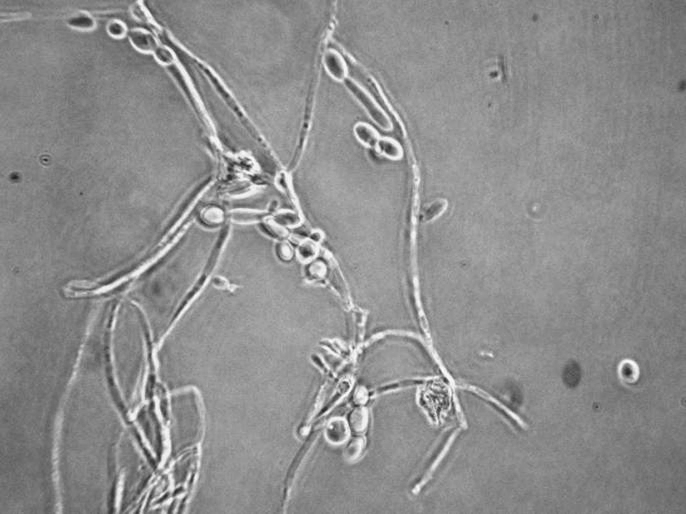
|
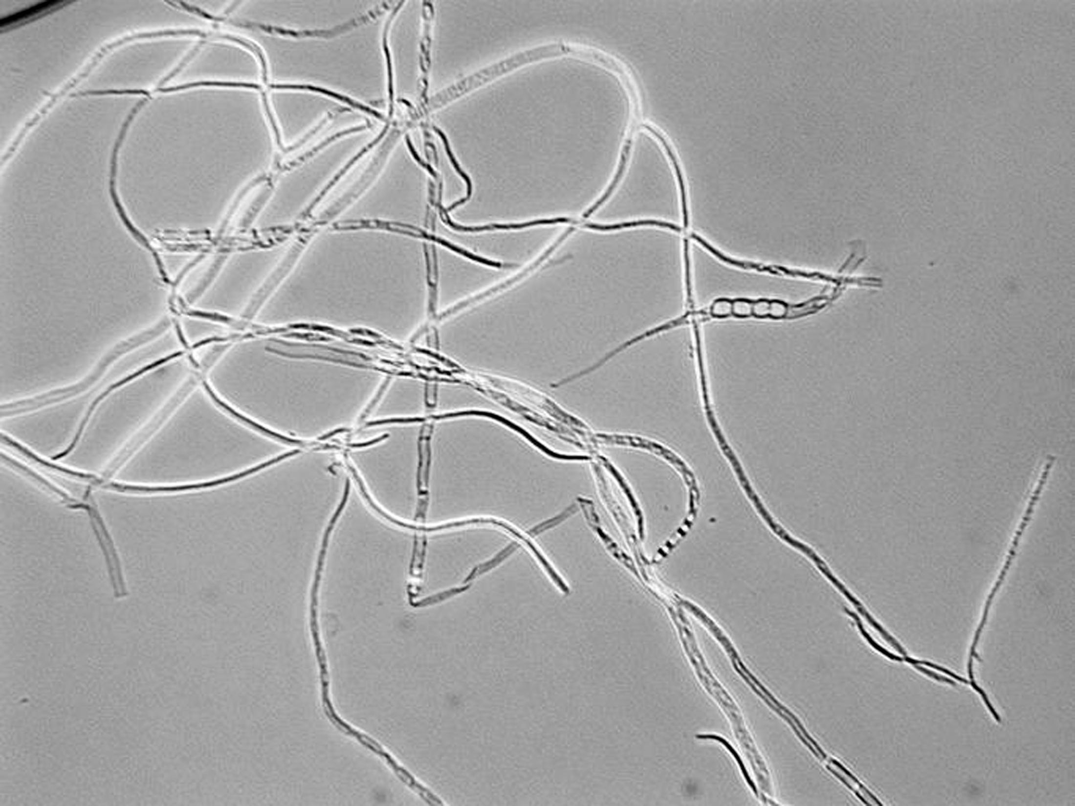
|
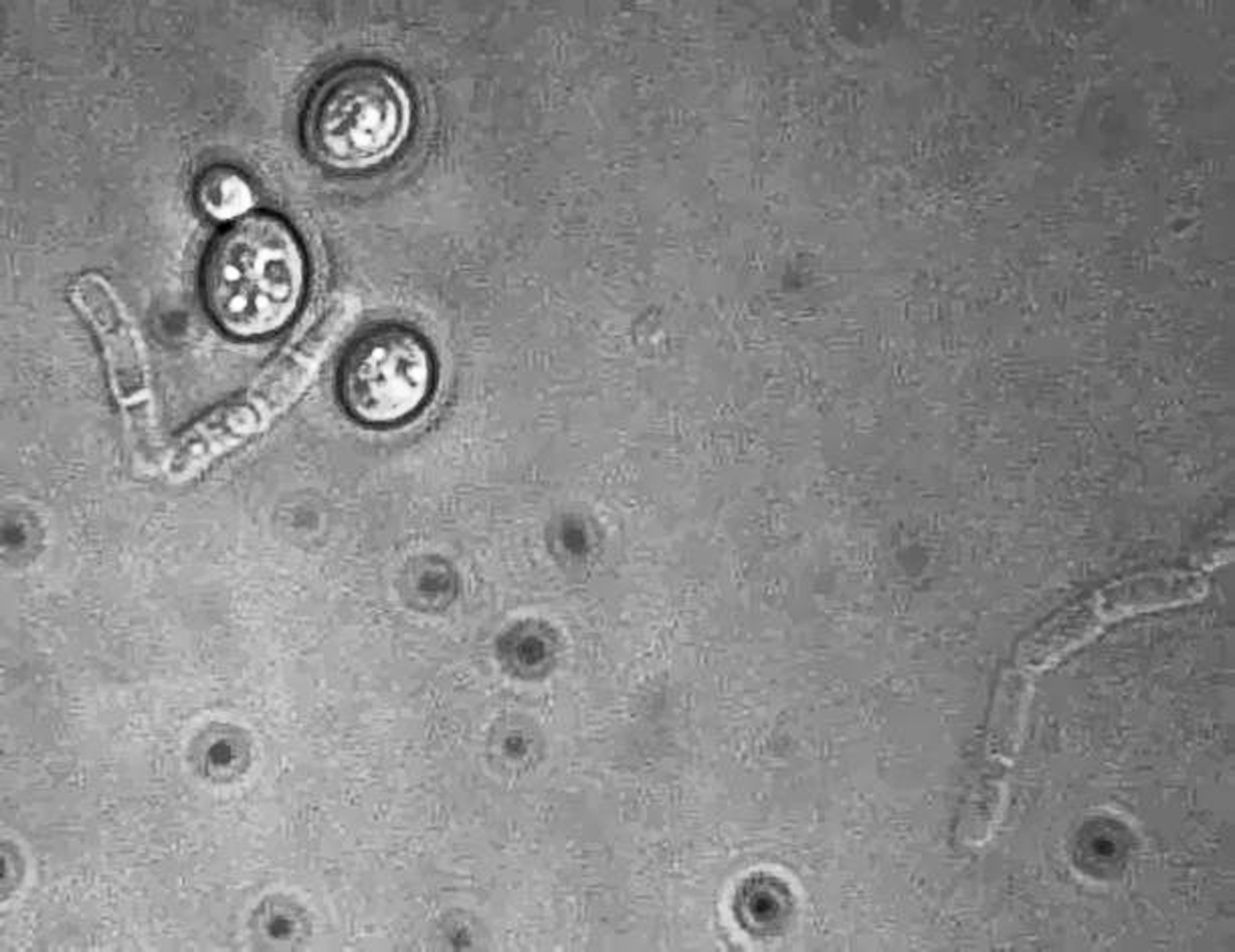
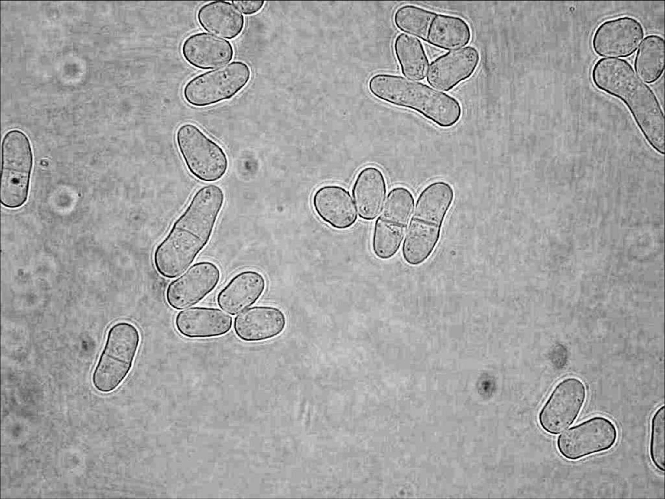
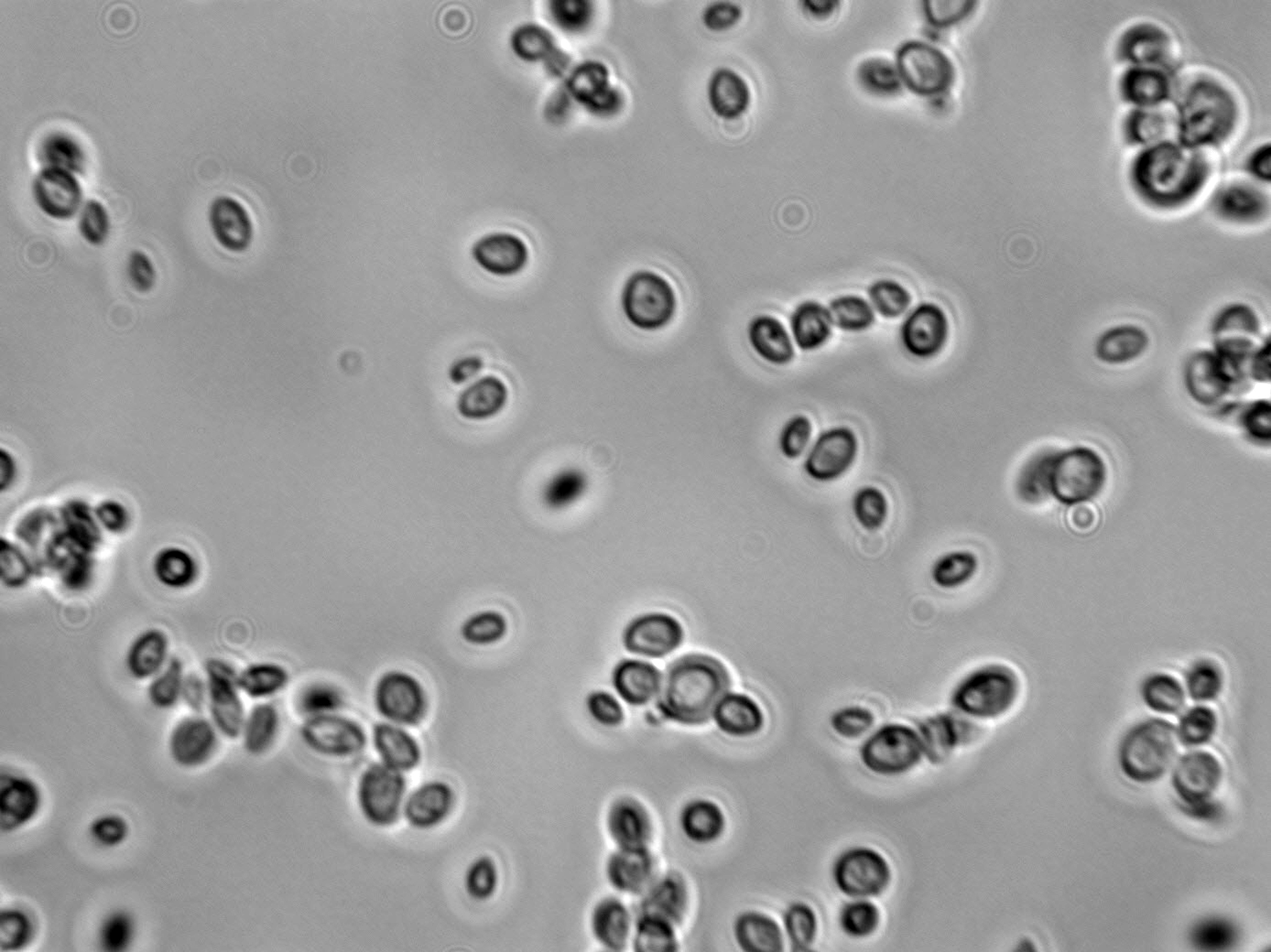
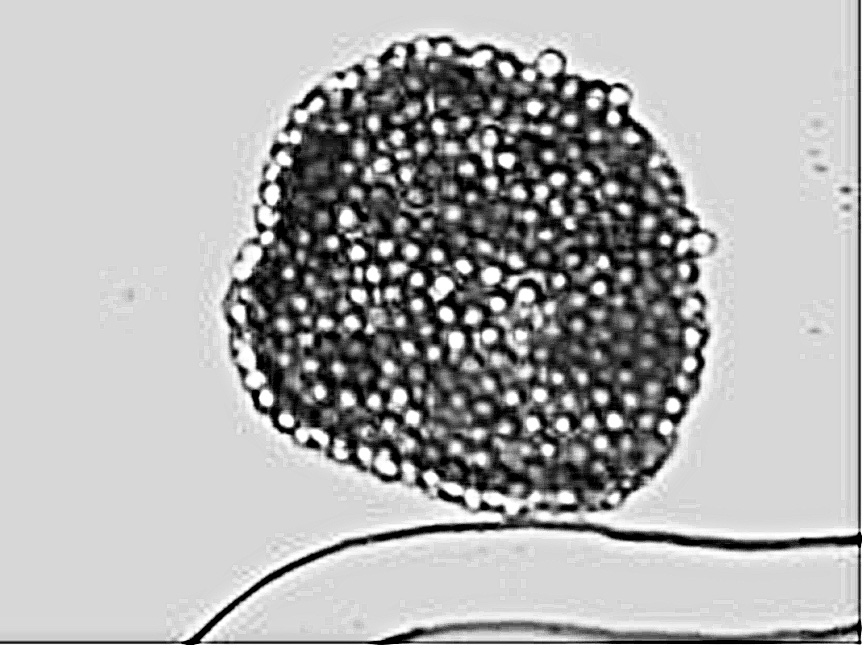
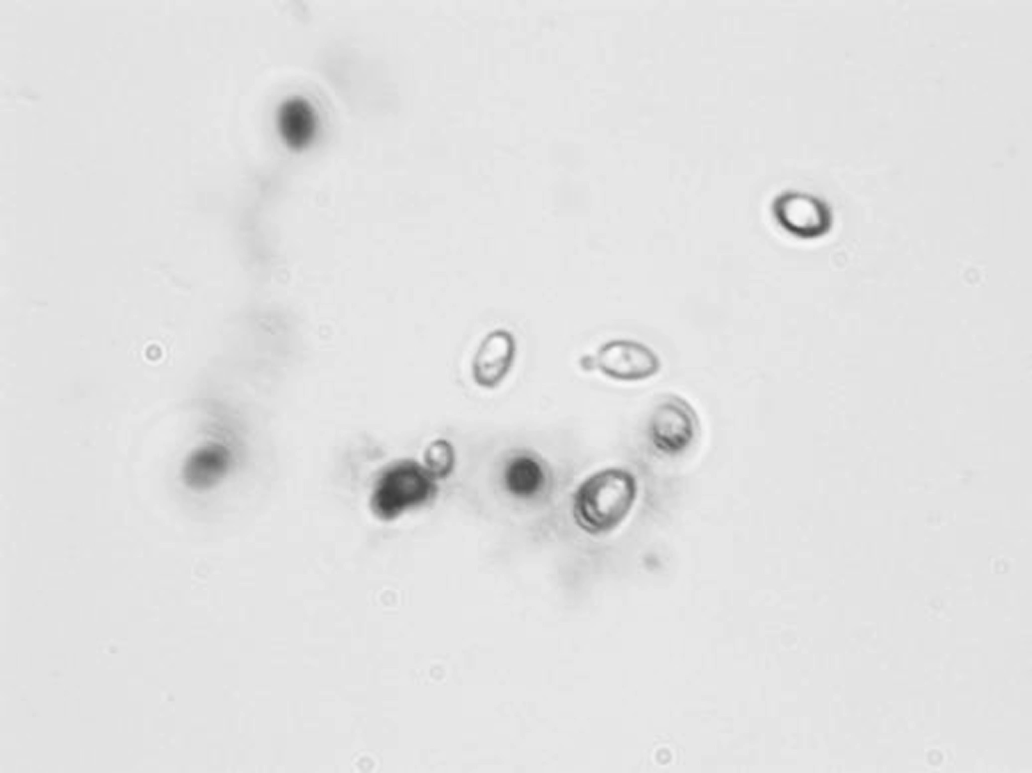
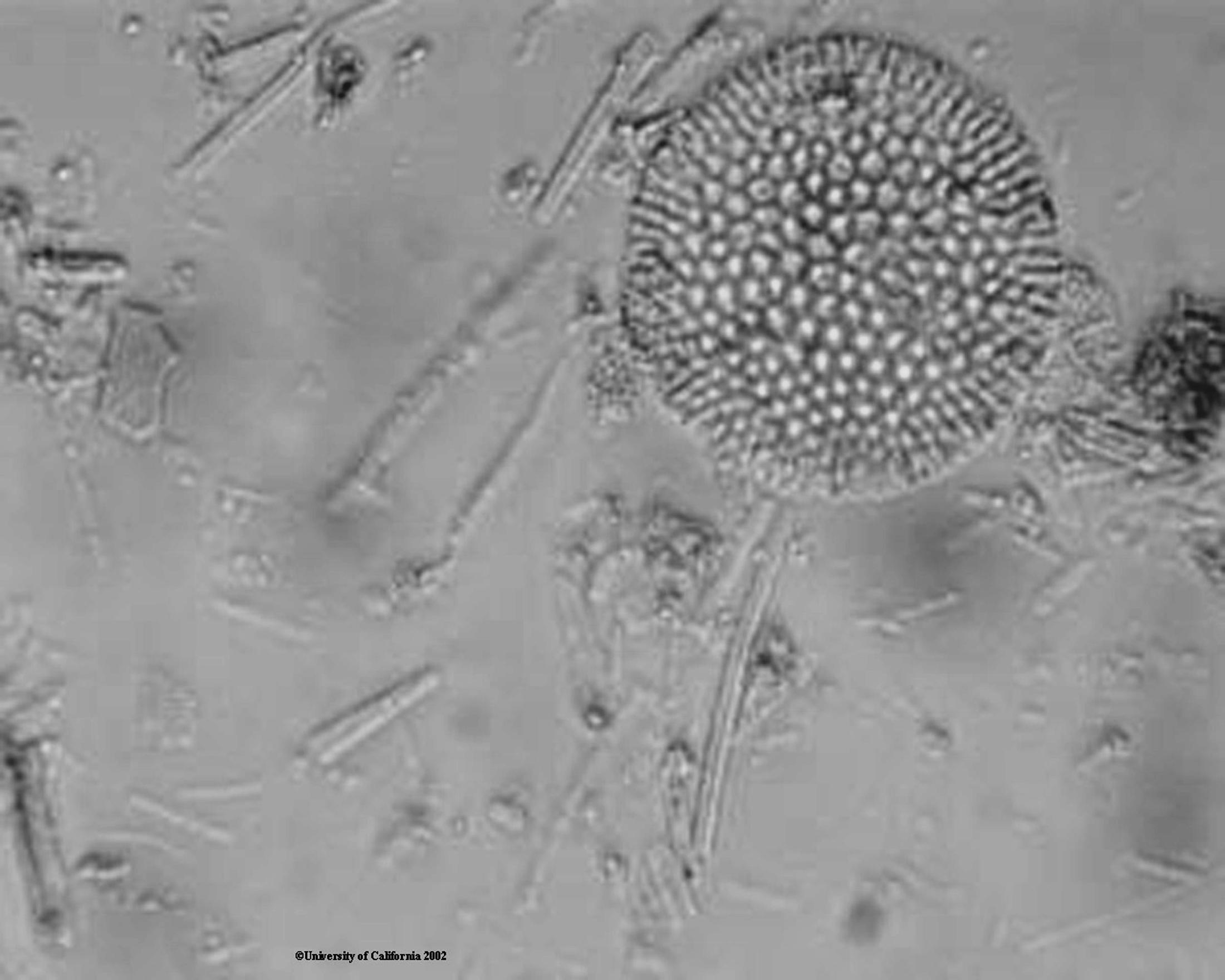
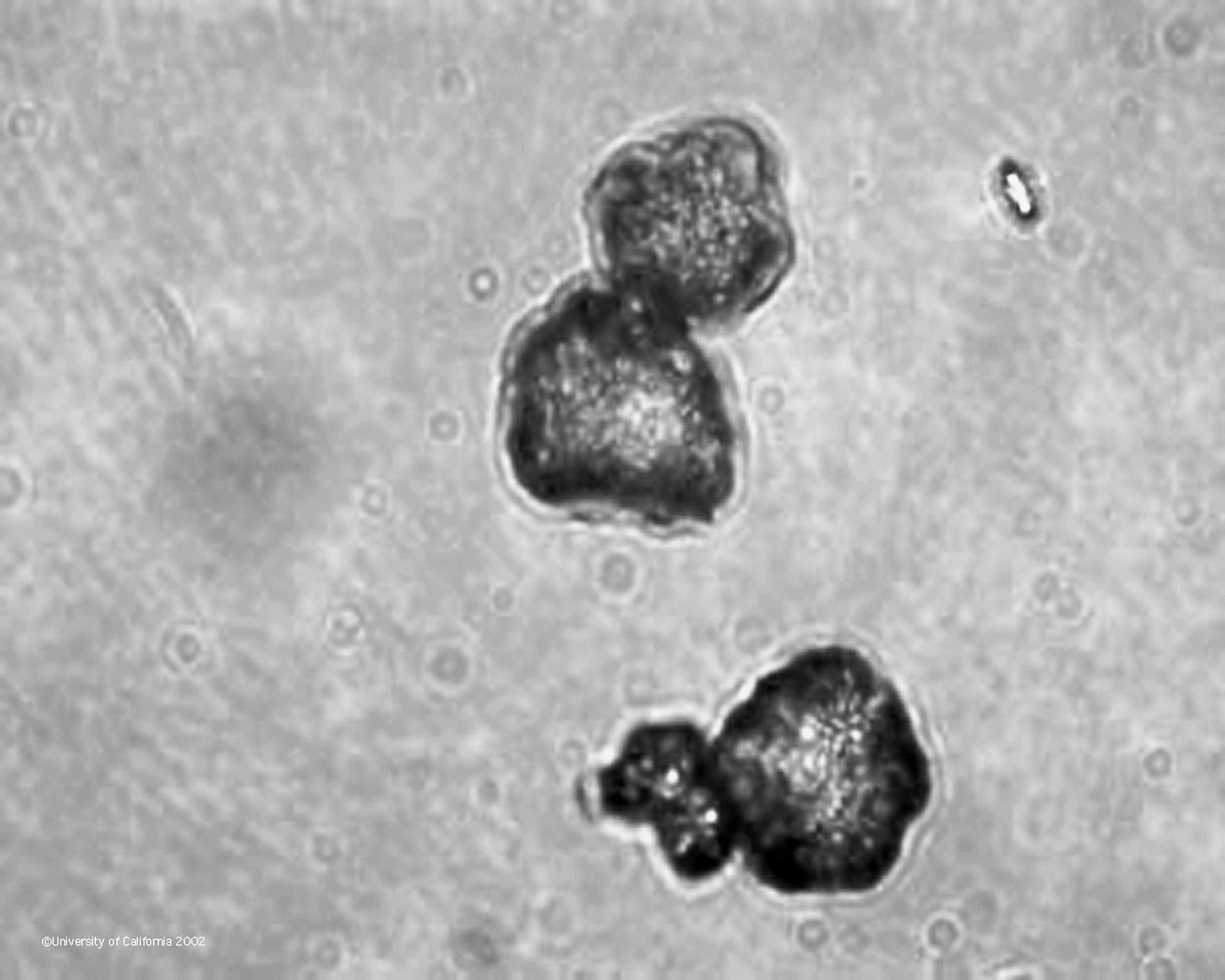
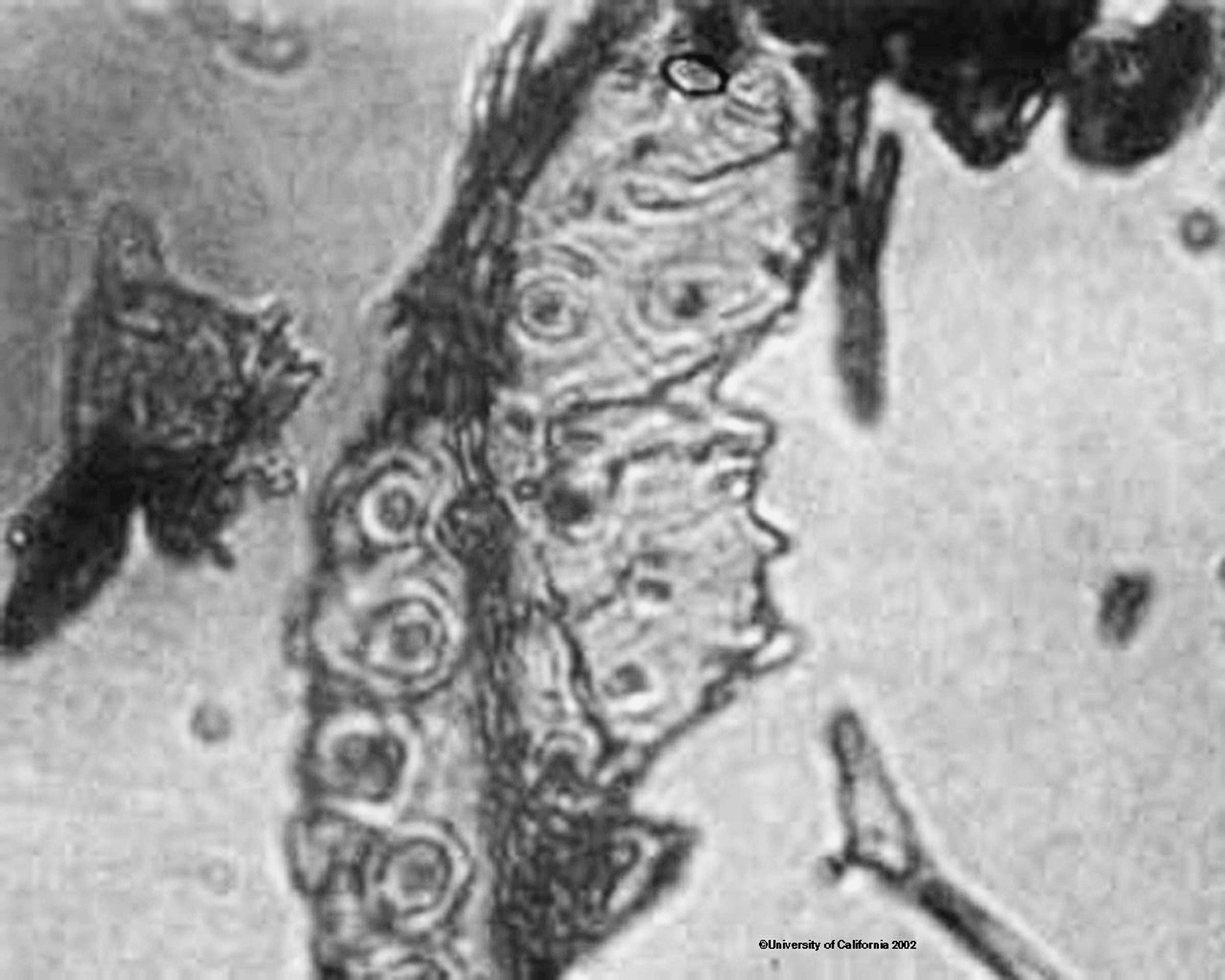
Another instance where it can be difficult to determine if you are seeing yeast or mold is when the mold has formed spores and you see loose spores. Below are some photos of yeast and mold spores for comparison.
|
|

|
|
|
|
Counting viable and non-viable yeast: (See also “Cell Counting – Total and Viable”) A microscope and a few simple tools can be used to give you an idea of the cell number and viability in your Saccharomyces inoculum. A simple counting chamber used to count blood cells or sperm can be used to determine the number of cells in a given volume and methylene blue dye can be used to determine the percent viability of an inoculum. Below is a photograph of the grid as you would see it under the microscope.
The image above is at 100x magnification and the cells shown are yeast cells. The square bounded by the 3 bright lines is 0.04 mm square, 25 of them will be 0.1 mm square or 0.1 ml in volume. Count enough squares to give you about 100 cells and then divide the number of cells by the number of squares counted, multiply by 25 squares in 0.1 ml and again by 10 to give you the number of cells in 1 milliliter. You must also take into account any dilution that you made of your inoculum by multiplying by the dilution factor to give you the number of cells in your original sample.
Methylene blue can be used to estimate the percent viability of the cells in your inoculum. You can purchase a methylene blue solution at the correct concentration to do your count. Prepare a microscope slide with 5 ul of dye and 5ul of your solution. Count 100 cells keeping track of how many are blue and how many are clear. The blue cells are are not viable, they cannot pump the dye back out after it penetrates their cell wall, and the clear cells are viable. The time the cells are in the dye is important. Try to count the cells at about 5 minutes after mixing the dye and the cell suspension. If you count to soon the dye may not have entered all the cells. If you wait too long some of the viable cells may no longer be able to pump the dye out and may become non-viable. The image below shows the same field of view at 2, 5 and 10 minutes. (See also “Methylene Blue Staining”)
|
|

|
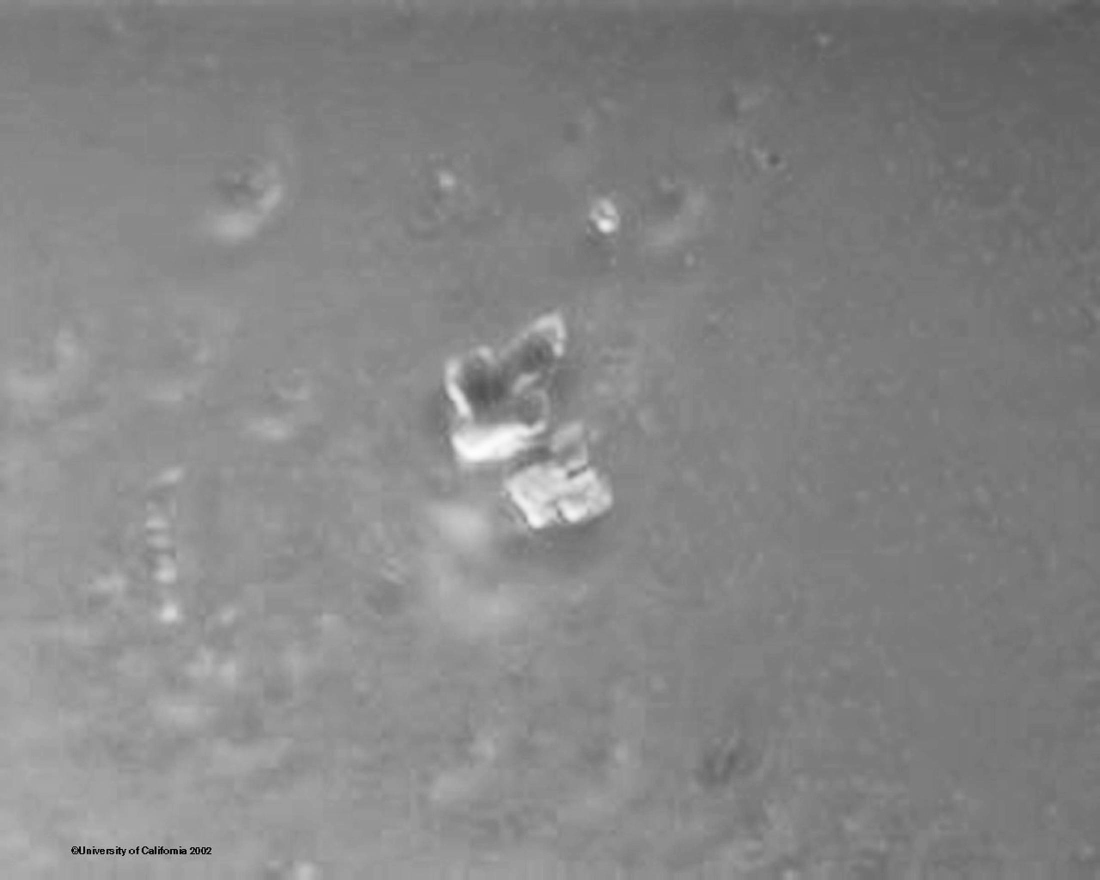
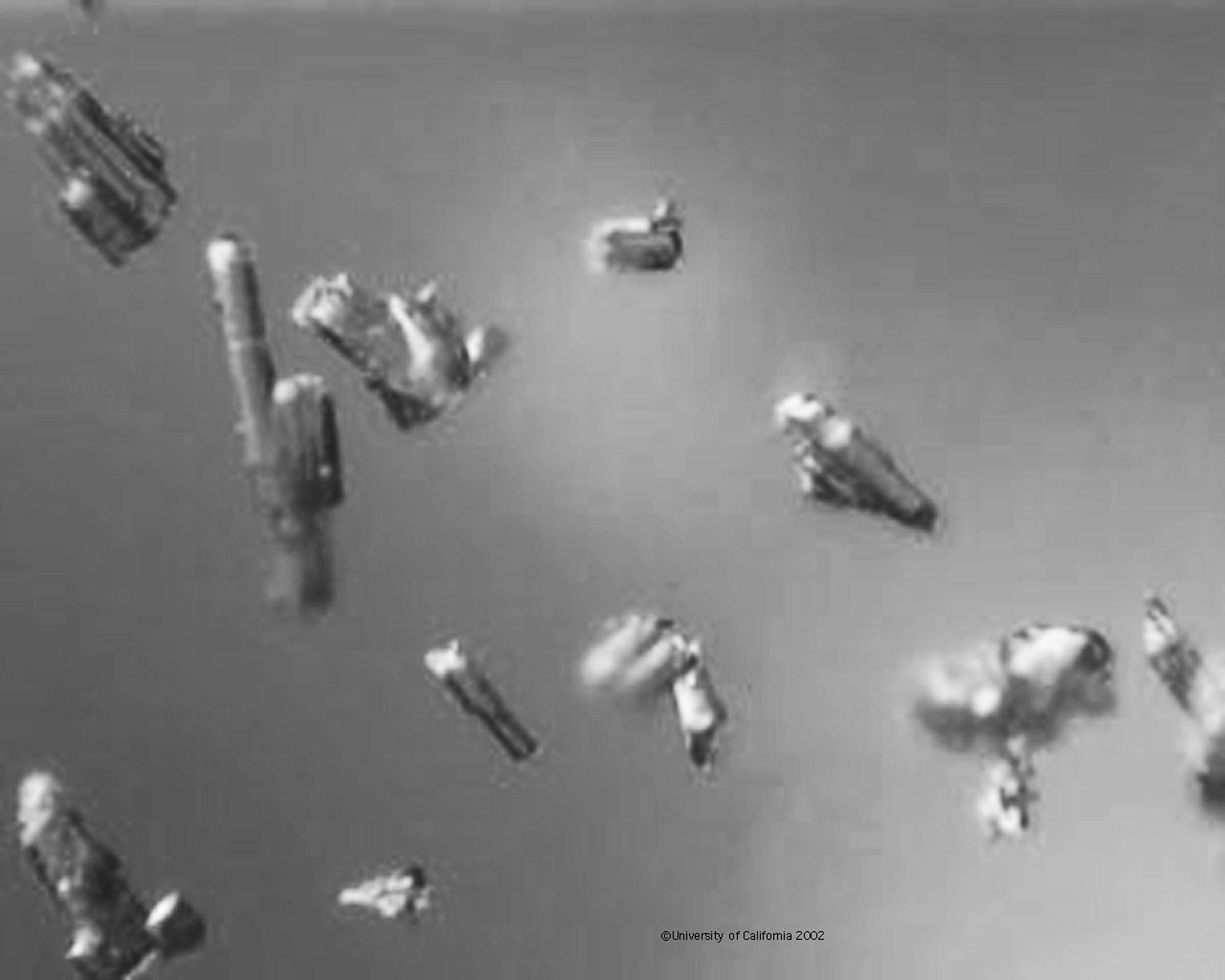
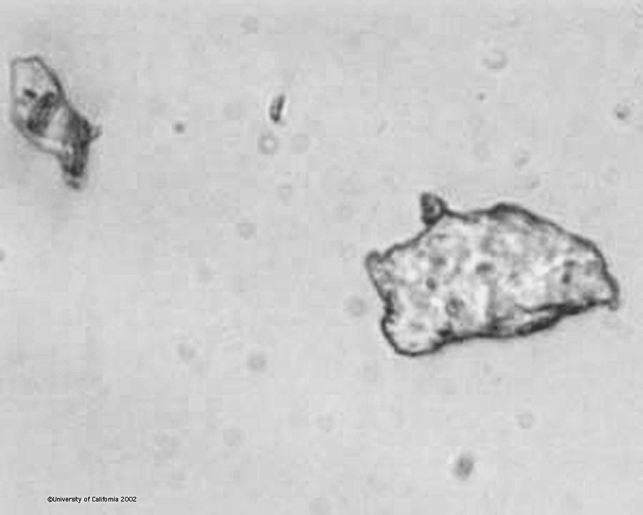
Distinguishing living cells from debri: The biggest clue to what is a biological organisms and what isn’t is symmetry. Debris tends to by asymmetric while living organisms are symetric. Of course many things that you will see while looking under the microscope are symetrical and not living, such as air bubbles, crystals, and dead cells. Crystals are easily identified by their geometric shape and sharp angles. Bubbles can be more difficult because they are rounded but they lack any internal structure and can be any size. They also tend to grow as the slide dries out. Dead cells can be harder or impossible to distinguish from live ones. Much of the debris in must is plant cells and will have cells that are joined together but ragged at the edges. Other particles you will see are often things that have been added to the wine such as fining or filtering agents. Some of these agents were once living and may be mistaken for living cells, such as the fossilized diatoms that make up diatomacious earth (DE). Below are some photographs of things you might see in fermentations and wine that are not alive.

|

|
|
|
|
|
|
|